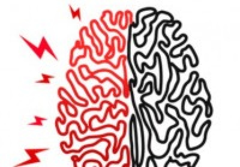
Cerebrovascular Accident (CVA or Stroke)
Over the last decade, EMS has seen a huge push in the industry to rapidly identify and treat the signs and symptoms of strokes. Similar to how materials on early recognition of cardiac arrest and impending cardiac arrest patients has seen large advancements, the AHA is also educating the public on what to look for when someone is having a stroke. The goal of this study guide is to supplement the education our EMTprep.com memberships provide that test EMT, AEMT, EMT-I, and Paramedic students.
We’ve said this in other posts, but it is worth repeating: the brain requires sugar, oxygen, and a pump to transport those items effectively. A cerebrovascular accident, also known as a stroke or CVA for short, is a sudden change in neurologic function caused by a disruption or sudden alteration in cerebral blood flow. Cerebral blood flow (the amount of blood being delivered to the brain) and cerebral perfusion pressure (MAP – ICP) are very closely related. With an increase in MAP or ICP, we will see a decrease in CPP, and thus a decrease in CBF. This relationship causes a sequence of events to occur that damages, and can ultimately kill, brain tissue.
Nationwide, strokes are the third leading cause of death. When blood flow to the brain is disrupted, a patient may experience significant changes in mental status, consciousness, and vital sign alterations such as; heart rate, respiratory rate, and blood pressure. With strokes may come an increase in intracranial pressure. (For more information on increases in intracranial pressure, click HERE to watch our video). Cells of the brain become injured and start to die if blood flow to the brain is impaired for longer than 10 to 20 minutes. Risk factors associated with strokes are carotid artery disease, cigarette smoking, obesity, family history, previous stroke, atrial fibrillation, cardiovascular disease, diabetes, and hypertension.
How the Brain Gets Supplied with Blood
Blood flow to the brain is provided by two arteries; the internal carotid artery and the vertebral arteries. The frontal lobes, anterior parietal lobes, and the anterior temporal lobes are supplied by anterior circulation which is provided by the internal carotid arteries. The brainstem, cerebellum, and posterior parts of the cerebrum are supplied by posterior circulation, which is supplied by the vertebral arteries.
Brain Statistics & How it Compensates
The brain consumes 20% of total oxygen in the body and 20% of total cardiac output when functioning normally. This is why it is so important to keep a constant supply of blood flow to the brain. In the event of a blockage of the anterior or posterior circulation, there is a system, termed the Circle of Willis, that allows perfusion to continue. The internal carotid arteries enter the cranial cavity and bifurcate into the anterior cerebral artery and the middle cerebral artery. The anterior cerebral artery is joined by an anterior communicating artery which forms the Circle of Willis. If the Circle of Willis becomes blocked, cerebral perfusion is limited to vessels in the dura mater and arachnoid matter.
The Two Types of Strokes
Ischemic Strokes
Ischemic strokes account for 80% of the strokes seen around the world. An ischemic stroke occurs when one of the arteries in the brain is blocked by either a thrombus or embolism. (A thrombus is a clot that forms at the blockage site. An embolism is a clot that forms elsewhere in the body, then travels to the blockage site). When blood flow is blocked (for either of the reasons listed above), all areas of the brain that are no longer getting perfused will stop functioning. This will manifest itself as deficits in the body region controlled by that area of the brain. The majority of ischemic strokes are caused by a thrombus created by atherosclerotic plaque formation in the cerebral arteries. These patients usually have a long history of cardiovascular disease. A lacunar stroke is a type of thrombotic stroke that occurs in the small vessels of the brain beyond collateral circulation. These types of strokes are usually devastating. Embolic strokes can be caused by air, fat, amniotic fluid, foreign bodies, tumors, bacteria, and fungus. Atrial fibrillation is a common cause of embolic strokes. A mural thrombus is a clot that forms in the atrial wall. The clot then breaks off the atrial wall, becomes an embolus, travels through the cardiovascular system where it becomes lodged in a cerebral vessel, and begins the stoke cascade.
Hemorrhagic Strokes
A hemorrhagic stroke occurs when a vessel in the brain ruptures causing a brain bleed. Intracerebral hemorrhages account for the majority of hemorrhagic strokes and are caused by bleeding located within the brain tissues. Subarachnoid hemorrhage is caused by bleeding beneath the arachnoid membrane. Hypertension, existing aneurysms, and arteriovenous malformations are commonly seen in a patient’s history experiencing a hemorrhagic stroke. Patients of all ages can experience hemorrhagic strokes. Trauma, ruptured aneurysms, and arteriovenous malformation are all causes of hemorrhagic strokes in the younger population.
Whether the patient is suffering from an ischemic stroke or a hemorrhagic stroke, time is crucial. In fact, people often say, “time is tissue.” The more time the stroke is allowed to run its course, the more brain tissue it damages as a result. Returning perfusion to the ischemic part of the brain is crucial in determining how the patient will recover. If reperfusion occurs before cell damage occurs, the patient can recover with minimal neurologic deficits. The “windoof treatment” is a term that relates to getting the patient fibrinolytic treatment within three hours of the onset of symptoms. Some facilities have extended the window to six hours. Time is brain when treating a stroke patient. As a paramedic, it is very important to answer the question “When did the symptoms start”? If this question cannot be answered, the patient automatically becomes ineligible for fibrinolytic treatment due to the risks associated with being outside the window of treatment. Bypassing a community hospital to go to a specialized stroke facility is generally a good idea when the patient is in the window of treatment. (As always, we at EMTprep.com cannot stress enough that you should always follow your local protocols).
Signs and symptoms associated with an ischemic stroke vary depending on if it is a stroke originating from a thrombus or embolism. Thrombotic strokes may cause hemiparesis or hemiplegia and symptoms may come and go, or slowly worsen with time. Progression of the stroke may take minutes to 3 days. An embolic stroke is associated with rapid severe symptoms without symptoms worsening or improving. Hemorrhagic strokes generally occur more rapidly and severely than ischemic strokes. Patients will often complain of a sudden onset of a headache and describe it as the worst headache of their life. Vomiting, neurologic deficits, visual disturbances, unconsciousness, and seizures are all signs and symptoms that may be seen with a hemorrhagic stroke. Signs of increasing intracranial pressure can be seen by unilateral pupil dilation, nausea /vomiting, and vital sign changes. Cushing’s triad is a late and severe sign seen with hemorrhagic strokes. This indicates brain herniation and is seen in the vital signs. Hypertension with a widening pulse pressure, bradycardia, and abnormal breathing patterns are signs of Cushing’s triad.
Assessing a Patient for Stroke Symptoms
The Cincinnati Prehospital Stroke Scale is a tool used by paramedics to help assess the presence of a stroke. This looks for the presence of facial droop, motor weakness, and/or aphasia. First, ask the patient to smile to determine if both sides of their face move well. If one side of their face doesn’t move and facial droop is noticed, this is an abnormal finding. Next, have the patient extend their arms out at a 90-degree angle with their eyes closed. If one arm falls before 10 seconds, this is an abnormal finding. If both arms move at the same time, or both arms do not move at all, this is a normal finding. Finally, ask the patient to say a simple saying such as “You can’t teach an old dog new tricks,” to assess for aphasia. If the patient repeats the saying clearly and correctly, this is normal. If the patient has slurred speech, uses inappropriate words, or can’t speak at all, this is abnormal. If one element of the Cincinnati Prehospital Stroke Scale is abnormal, there is a 72% chance they are experiencing a stroke.
Treating Stroke Patients
Treatment patients for a stroke depends on getting them to the hospital in an expedited fashion, especially if signs show the patient is suffering from an acute stroke. It is important to activate a stroke alert to notify the receiving hospital that a stroke patient is en route to their facility. Just like any patient encountered in the field, the primary concern revolves around the patient’s ABC’s. Depending on the location of the stroke, the patient may lose their ability to swallow or breathe effectively. In this instance, as a paramedic, securing a patent airway is the primary concern. If pharmacologically assisted intubation is necessary, consider using lidocaine to prevent increasing ICP. There is a decent amount of evidence that suggests Etomidate does an adequate job at reducing ICP. Apply oxygen as needed, put the patient on a monitor, establish IV access, and obtain a 12-lead. A saline lock or a TKO drip rate at 30 ml/hr is sufficient for a stroke patient. Do not give large volumes of fluid unless hypotension is present. Check the patient’s blood sugar. Only give dextrose if the patient presents with a CBG below 60 mg/dl. Keep in mind, a stroke patient may not be able to communicate due to the neurologic deficits, but the patient may be fully aware of what is going on around them. Keep professional, provide a high standard of care, and get them to the appropriate receiving facility in a safe, but expedited fashion. (As always, we at EMTprep.com cannot stress enough that you should always follow your local protocols).
At EMTprep.com we do our best to ensure that our content is constantly reviewed and updated when needed. We also make every effort to ensure that our resource posts reflect the content in our quizzes, tests, study guides, and other study materials. Be sure to check out one of our memberships whether you’re trying to study up for a certification exam, employment exam, or you just want to brush up on your knowledge.
- Dozens of courses and topics
- State-specific requirements
- We report to CAPCE in real time


