Atrioventricular Blocks Study Guide
Arguably the most difficult thing to remember in ECG class is Atrioventricular (AV) Blocks. The goal of this study guide will be to clarify any information you find confusing AND provide you with an easy way to remember the different types.
Let’s go back to some A&P. We learned that the AV junction is the area that links the atria and the ventricles. Should a person experience a problem with conduction in the AV node itself, the bundle of His, or the His-Purkinje system, they will present with an atrioventricular block.
We get asked many times throughout the year, how do you differentiate between the 4 types of AV blocks effectively? The answer is typically found in the PR-Interval. If you want to know where the block is occurring, look to the QRS width and heart rate. We will explore this a little more below.
If we’re honest with ourselves, sometimes AV blocks require intervention from EMS, and other times they don’t. When confronted with one on your ECG strip, ask yourself what type of AV block you’re dealing with, how fast (or slow) is the rate, and how is your patient presenting (symptomatic or non-symptomatic)?
First-Degree AV Blocks
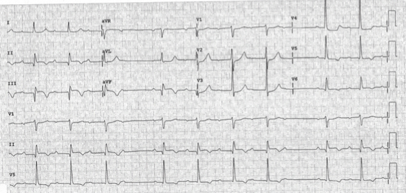
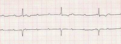
- A first degree AV block will look nearly identical to normal sinus rhythm. However, there is a delay at the AV node that still allows all impulses to be transmitted through. This delay shows up on our ECG strip as an elongated PR-interval. How long is long here? A PR-Interval greater than 1 big box, 5 small boxes, or .2 seconds.
- Characteristics
- Regular Rhythm
- Variable rate, but the atrial and ventricular contractions are equal
- One P-wave to each QRS
- A constant but elongated PR-Interval of >.20 seconds
- QRS width is generally normal
- Etiology
- An increase in vagal tone
- Myocardial infarctions
- Prescription medications (Digitalis, Amiodarone, or any of the Calcium/Beta-blockers)
- Idiopathic
Second-Degree Type I AV Block (Mobitz 1 or Wenkebach)
- This AV block is characterized by its lengthening PR-Interval until a P-wave is generated with no subsequent QRS spike. This is commonly referred to as a ‘dropped beat.’ If we think back to our A&P, you’ll remember that in 90% of the human population, the RCA supplies the AV node. So as you can imagine, if your patient occludes their RCA, they could very well present with a Second-Degree Type I rhythm
- Characteristics
- A regular atrial rhythm with an irregular ventricular rhythm
- The rate will vary
- There will be more P-Waves than QRS spikes
- The PR-Interval will become increasingly longer until one p-wave is not conducted, leading to the dropped beat
- The QRS width will be greater than .12 seconds or 3 small boxes
- Etiology
- An increase in vagal tone
- Inferior myocardial infarctions
- Cardiac prescription medications
- AV nodal insult
Second-Degree Type II AV Block (Mobitz 2)
- With this type of AV block, you have the SA node successfully creating P-waves, however, due to the location of the AV block, only one impulse actually makes it through to create a QRS spike. This block will originate in one of two places, either the Bundle of His or at the bundle branches. This is a critical finding and very often these patients will deteriorate into a third-degree AV block
- Characteristics
- Regular atrial rhythm and an irregular ventricular rhythm
- The rate is variable
- The P-waves are normal in appearance, however, there are more P-waves than QRS spikes
- The PR-interval can be either normal or prolonged, but it remains constant
- The QRS will be either narrow or wide
- Etiology
- Hypoxemia
- Myocardial infarctions
- Cardiac medications
- AV nodal insult
Third-Degree AV Block
- The key to remember with third-degree heart blocks is that it is a very serious finding in your patient. The heart has no connection between atrial and ventricular contractions and that is why it is also described as “complete heart block.” Typically, you will see P-waves at a rate of 60-100 beats per minute and QRS spikes at a rate of 20-60 beats per minute.
- Characteristics
- Regular atrial and ventricular rhythms, yet the two are disjointed
- Sinus P-waves are present with no relation to the QRS
- The PR-interval varies greatly as noted above
- The QRS will either be wide or narrow
- Etiology
- Acute myocardial infarction
- Cardiac medications
- Ischemic heart disease
- AV nodal insult
Quick and Easy Reference Chart
|
- Dozens of courses and topics
- State-specific requirements
- We report to CAPCE in real time


